显示标签为“Blog”的博文。显示所有博文

2013年9月28日星期六

Dog Ear Infections blog


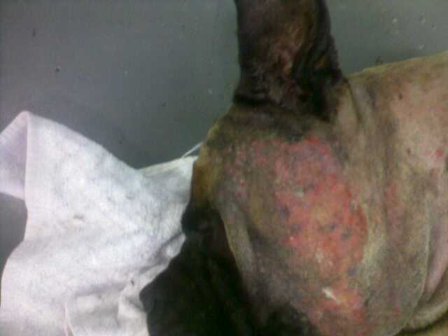
Dog ear infections can become severe if left alone, and even cause other problems.  One common complication of dog ear infections is an aural haematoma.  An aural haematoma is like a big blood blister in the ear of a dog.  It often results from the dog scratching at its ears and shaking its head, as it does with an ear infection.  Sometimes, it occurs from another injury, and sometimes there isn’t an obvious cause.
Aural haematomas start off small, often only 1/4 of the ear.  They frequently grow, especially if the dog keeps shaking its head.  Aural haematomas are normally treated surgically under anaesthetic by removing the blood and stitching the two sides of the ear flap together.  WIthout these stitches, the blood would just fill the ear again.

Its very important to check the ears thoroughly and rule out an ear infection as if this isn’t treated properly there may be further complications.

This article on aural haematomas shows a good picture of a large haematoma on a dog.


Aural haematomas in dogs – ears


An aural (ear) haematoma is a collection of blood or serum, and sometimes a blood clot within the pinna or ear flap. This blood collects under the skin and causes the ear flap to become thickened. The swelling may involve the entire ear flap or it may involve only a small area.


Normal Canine EarNormal Canine Ear


How does an aural haematoma occur?


Aural haematomas usually occur as a result of local irritation to some part of the ear. When something irritates the ear canal, a dog is likely to respond by scratching or shaking the head. Excessive shaking causes blood vessels to break, resulting in bleeding. An understanding of the ear’s anatomy makes the sequence of events more logical.


Understanding ear anatomy


The ear flap is composed of a layer of skin on each side of a layer of cartilage. The cartilage gives the ear flap its shape. Blood vessels go from side-to side by passing through the cartilage. Violent shaking causes the vessels to break as the skin slides across the cartilage.


How are aural haematomas treated?


The first aim of treatment is to drain the haematoma to relieve the pressure and pain associated with the build up of fluid within the ear flap. This is achieved under general anaesthesia where either a single incision or multiple small biopsy holes are made on the inner surface of the ear. The blood is drained and the ear flushed to remove any remaining blood clots. These holes are left open to allow continued drainage of fluid whilst waiting for the ear flap to heal.


Reattachment of the ear cartilage is encouraged with the use of multiple sutures placed through the ear flap (with or without the use of a support to maintain the normal architecture of the ear) and these sutures are left in place for 3 weeks. The specific method used will depend on the size, age and position of the haematoma.


The second major aspect of treatment is to work out why the haematoma formed in the first place. As mentioned, above, any reason that causes the dog to shake its head can result in the formation of an aural haematoma. Some things which can cause this include:


  1. Grass seed or other foreign body lodged within the ear canal.

  2. Ear infection.

  3. Allergies resulting in an itchy ear, scratching and shaking head.

  4. Fly bites to the tips of the ears.

  5. Immune mediated disease.


It is essential that the cause of the problem be identified and treated if possible. If a foreign body is found, it is removed. If an ear infection is identified, the ear canal will be thoroughly cleaned during anaesthesia and appropriate medical ointments or medications will be dispensed.


Unfortunately, it is not always possible to identify a cause, or it is difficult to manage the underlying cause (eg allergies). In these cases, another aural haematoma may form in the same ear or in the other ear and management may require long term medications.


Aural haematoma - showing swelling of the pinnaAural haematoma – showing swelling of the pinna


Once my dog is treated will I need to bring him/her back to the vet for further treatment?


The sutures will need to be removed 3 weeks after surgery. At this time, a haematoma is usually healed. If an infection is also being treated your veterinarian will also check to make sure that the infection is gone. It is vitally important that the infection is successfully treated to prevent further head shaking which may result in further haematomas.


What happens if your dog does not have surgery?


If a haematoma is left untreated the blood in the ear flap will separate into serum and a clot and will gradually be absorbed over a period of 10 days to 6 weeks. This is an uncomfortable time for your dog and unfortunately some scarring will take place during this process. It also causes a deformity of the ear flap resulting in a “cauliflower ear” which may cause further problems.


Do your dogs ears smell? Dog ear infections are very common and often the first sign is a smelly, itchy or red ear.  If the infection is worse, there is often pain and a discharge which may be foul smelling.  Learn how to identify these problems early and treat dog ear infections before they turn into something more serious.

Dog Vet


Dog ear infections most commonly start as infections of the outer ear, but if neglected they can spread into the middle and inner ear.  this often involves damage to the eardrum.  For this reason, its vital to get your vet to make sure the ear drum is intact before you consider using chemicals in the your dogs ears.  This article “Otitis externa, otitis media, and otitis interna   “  gives a good explanation of how a simple dog ear infection can turn into something much more serious.
Dog Vet


Otitis externa, otitis media, and otitis interna     
Ear infection, inflammation of the external ear canal, middle ear infection, inner ear infection Affected Animals 
Dogs and cats. Cocker spaniels and other long-eared or floppy-eared breeds may be more predisposed to developing infections.


Overview
An ear infection, or otitis, is an inflammation of the outer, middle, or inner ear canal. Most frequently, a dog will develop otitis in the outer ear that may worsen and spread into the middle ear. Once in the middle ear canal, the inflammation can move into the inner ear — or, in cases in which the otitis has originated in the middle ear, the infection can instead progress outward to the external ear.


Otitis can be caused by a tremendous array of factors, including fleas, excess liquid in the ear from swimming, autoimmune diseases, skin parasites, and excess wax production. Generally, dogs that develop ear infections have reddened ears that are painful to the touch and ooze a foul-smelling liquid. A punctured eardrum is not an uncommon result of a middle ear infection, and the more severe cases of otitis can lead to partial deafness, lack of balance, nausea and vomiting, and problems with the nerves of the eye.               Clinical Signs
Clinical signs can vary depending on the severity and location of the infection, but typical symptoms of otitis include pain and erythema of the pinna, head shaking and scratching of the ears, and purulent and malodorous exudate coming from the ear canal. The animal may tilt the affected ear downwards and may roll or lean to the affected side. If both sides are affected, the animal may be deaf or ataxic. Some animals may be nauseated, vomit, and have Horner’s syndrome. Any neurological clinical signs indicate significant middle ear or inner ear disease.  Symptoms
Ears that are red, painful to the touch, and produce a foul-smelling discharge are symptomatic of otitis. Typically, a dog with an ear infection will scratch and shake the ears or may tilt the affected ear downwards. Animals that are affected more severely may show some neurological signs such as rolling or leaning to one side. If both ears are affected, the animal may be deaf or off balance and uncoordinated. Some animals may be nauseated and vomit. Also, some dogs may get a condition called Horner’s syndrome in which the pupils are sized differently and the nictitating membrane is raised. This condition indicates that a nerve has been affected by the inflammation from the middle ear. Any neurological clinical signs indicate significant middle ear or inner ear disease.  Description
Typically, ear infections begin with otitis externa and then progress deeper into the canal to the middle ear. When the inflammation in this region of the ear is chronic, the eardrum may rupture and the infection may spread to the inner ear — or, the infection may begin in the middle ear and progress outward to the external ear. Of the three types of otitis, infections in the inner ear are often the most severe and can lead to partial deafness and neurological problems. In serious cases of otitis, the skin begins to form into folds in which the infection can become trapped, making cleaning and use of topical treatments very difficult. In addition, in cases of otitis interna, the skin will secrete more wax and debris that allows yeast and bacteria to overgrow, causing further disease. Severe inflammation leads to permanent skin thickening, mineralization, and narrowing of the ear canals. Once this occurs, the only viable treatment will be surgical removal of part or all of the ear canal.                 Diagnosis
The veterinarian will make a diagnosis based on the clinical signs, physical exam findings, and through the use of several other diagnostic tools. One such tool is cytology, which involves taking a swab of the ears and looking at the material collected under a microscope for the presence of bacteria, yeast, mites, and other substances that might cause an infection. Bacteria and yeast are normally present in low numbers in all dogs’ ears, but a large presence will lead to an ear infection. Skin biopsies may be needed to determine any diseases such as an autoimmune disorder that could cause a skin abnormality affecting the ear. Skin scrapings may be needed to detect mites, tiny parasites that can infect the ears and cause skin diseases. X-rays of the skull can be used to examine the middle and inner ear for signs of disease. In addition, cultures of an infected ear help determine the presence and type of bacteria, as well as antibiotics that are appropriate for treatment. The majority of animals with ear problems have such pain in their ears that they cannot withstand having them examined, cleaned, cultured, or x-rayed without the use of general anesthesia.              Prognosis
With proper treatment, otitis externa usually will resolve within three to four weeks, although it may recur in certain animals. The prognosis is good for otitis media and otitis interna since medical treatment usually is effective. The key for long-term success is correcting or treating the underlying problem that led to the development of the otitis.   Transmission or Cause
Possible causes of otitis externa, which may lead to infections of the middle and inner ear, include excess moisture in the ears due to swimming or bathing; inappropriate treatment or cleaning of the ears such as may be caused by the use of cotton-tipped applicators; excess wax production in the ears; an underlying disease; or a foreign body that is obstructing the ear canal. Additionally, otitis externa can be caused by a host of parasites, including ear mites, mange, fly bites, and fleas. Allergies and illnesses such as autoimmune diseases may also lead to otitis. The most common cause of the development of otitis media and otitis interna is a bacterial infection resulting from otitis externa.   Treatment
After the cause of the otitis has been diagnosed, the veterinarian usually will perform a thorough cleaning of the ear canals while the dog is under sedation or general anesthesia. The use of topical medications, which are placed into the ear canal, is often very beneficial in killing yeast, bacteria, and mites. Oral medications also may be used in conjunction with other treatments to help kill bacteria, yeast, and mites. The veterinarian will teach the dog’s owner how to clean and medicate the ears properly to ensure successful treatment of otitis. The surgical correction of the ear canals may be necessary in cases of severe infection. One type of surgery, called a lateral ear canal resection, allows the ear to drain more easily, decreases the amount of humidity in the ear, and makes topical treatments easier to apply. A total ear canal ablation — a complete removal of the ear canal — is performed on dogs with severe, chronic ear disease that is nonresponsive to medical therapy.            Prevention
Prevention is best accomplished when the veterinarian is able to determine the underlying factors that can lead to development of otitis. Proper cleaning of the ears is also critical. A veterinarian can explain how to clean the ears, as well as how to apply any medications that have been prescribed to treat the otitis.


Dog Ear Infections vary form mild to severe and have many causes.  To your average owner, these all look the same, but your vet has the skills, experience and equipment to locate the cause and assess the severity of the problem.

Morris Animal Hospital has a great web page showing just what infected, dirty, hairy and blocked ears look like inside.

Very interesting stuff.


Dog Vet

Dermatology and Diseases of the Ear 
Otoscope Case Examples


A normal canine eardrum.  The eardrum is located 3 inches deep in the canal in large dogs.  Note the eardrum (green) and the small amount of wax adhered to the normal tuft of hair that frequently grows next to the eardrum (red).
 
Excessive amounts of hair can be normal in breeds such as the Poodle and Bichon Frise.  Sometimes the hair causes moisture and wax to be retained in the canal causing an ear infection.
 
This large wax plug located deep in the ear canal of a cat caused a good deal of discomfort.  It was removed under anesthesia with the video otoscope.


This Labrador Retriever had severe bilateral otitis and was in excruciating pain.  Note the copious amounts of discharge and wax which was subsequently cleansed from the canal with the video otoscope.
 
Polyps are sometimes the cause of chronic ear infections. Note how this large polyp is completely obstructing the canal.  Polyps cannot be diagnosed and adequately treated until the canal has been thoroughly cleaned.
 
This older cocker spaniel has likely had ongoing infections for years.  Note the hemorrhage and inflammation.  This photo was taken after a tremendous amount of wax and debris were removed. Her ear drum is ruptured but she has taken her first step toward full recovery.


A young female German Shepard named “Heidi” with a diseased canal lining and ruptured ear drum.  She later had surgery to reroute her ear canal which helped prevent future infections & relieved her pain.A springer spaniel with complete ear drum rupture & otitis media (middle ear infection).  Beyond the hair where the ear drum should be, the interior of the middle ear can be seen.  This dog needed antibiotics for 6 weeks to resolve her middle ear infection.After extensive flushing and removal of debris, a normal ear drum is finally visualized in the depths of this ear canal.  The catheter being used in this photo is only 1.5 mm in diameter.  It is impossible to clean ear canals to this extent without the use of the video ototscope.


Dog ear infections commonly recur.  People often blame this on antibiotic resistance or problems with their vet, but the main causes are actually failure to ensure the ear is clean (and stays clean afterward) and failure by owners to put ear drops in the correct location.  The wikipedia article on dog ear infections has this useful image describing where to put ear drops when medicating your dogs ear.


File:DogEarMedicationSW.JPG 


Dog ear infections are common, but there are many possible causes.  I love this article 8 Possible causes of dog ear infection because its simple, accurate, and straight to the point.


Dog Ear Infection


8 Possible Causes Of Dog Ear Infection


Dog ear infection, otitis externa, is a condition of the inflammation of the outer ear canal.


It is a common condition in pets. An estimate of 20% of the dog population is affected by this ear problem.


The structure of a dog’s ear canal differs from human. Human has a horizontal ear canal. A dog has a L-shape ear canal. It is easy for ear wax and moisture to be trap in the vertical section of the dog’s ear canal. Often, the blockage leads to a dog ear infection.


There are many reasons for ear infection in dogs. Knowing the cause of the problem comes in handy when you want to help your dog to get better.


Here are 8 possible causes of dog ear infection:


  • Floppy versus Erect Ears


Long floppy ears blowing in the wind makes a dog looks stylish and elegant. But those same floppy ears have a drawback. They block off light and reduce the air circulation within the ears.


Yeast and bacteria multiply quickly in a dark, moist and warm environment. The ear canal of a long, floppy-ear dog is the perfect place. Breeds like Basset Hound, Beagle and Cocker Spaniels often have this ear problem.


  • Micro Organisms


Bacteria and yeast are one of the most common factors in dog ear infection.


The most common bacteria associated with ear infections are Staphylococcus Intermedius, Proteus Mirabilis, and Pseudomonas Aeruginosa. Pseudomonas bacteria infections are very resistant and often notoriously difficult to control.


Malassezia Pachydermatis is the yeast found most frequently in association with ear infections. It can cause severe ear and skin infections.


A healthy dog has a balanced flora of yeast and bacteria in the ear canal. But when the ear environment changes due to excess moisture, allergies or hormone disorder, the yeast and bacteria multiply quickly and the ear becomes inflammed.


  • Parasites


Ear mites cause intense itching to your dog. A dog’s ear that is infested with ear mites can have thousands of these tiny bugs crawling in the ear canal. As the mites feed and eat into the skin, the ear canal becomes sore and inflamed.


A dog trying to relieve the itch by scratching relentlessly, may cause further damage to the ear flaps.


  • Allergies


Underlying allergies sometimes manifest as dog ear infection. A dog can have an allergic reaction to its food, something it inhales, or something it comes in contact with.


It is important to address the allergy in addition to the infection so as to minimize recurring ear inflammation.


  • Foreign Bodies


Things that can get into your dog’s ear include insects, twigs, grass seeds, sand, water, pet’s fur … and the list goes on. The foreign object causes irritation. If it travels deeper into the ear, it can cause damage to the ear drum.


  • Hematoma


Sometimes a ear infection leads to hematoma in a dog. A dog scratching and shaking its head vigourously to get rid of an itch can break the blood vessels in the ear flap causing bleeding. The ear flap becomes swollen as blood collects within the ear flap. This can be very uncomfortable for the dog.


  • Hypothyroidism


Hypothyroidism is the condition where one has inadequate active thyroid hormone. It is the most common hormone imbalance of dogs. Hypothyroidism generally develops in middle aged or elderly dogs.


An imbalance in the thyroid hormone can also lead to dog ear infection.


  • Tumors


Ear tumors in dogs can be benign or malignant. They generally affect middle aged or older dogs. Ceruminous gland adenocarcinoma is the most common malignant tumor of the ear canal of dogs, especially cocker spaniels. Other tumors may include papillomas and squamous cell carcinomas.


Some tumors cause pain, ulceration, bleeding and secondary ear infections.


If your pet has dog ear infection, do you know what causes the ear problem?


It may be difficult for a pet guardian to accurately determine the cause of a dog ear infection. So it is advisable to bring your dog to the veterinarian. An otoscopic examination and some lab work would provide a diagnosis of the ear infection.


Once you know the cause of the dog ear infection, you or your vet would be able to apply the appropriate treatment options.


Ear infections in dogs range in severity from mild to severe.  This article on ear infections highlights why its so important to seek veterinary advice.  I’m sure if you have researched dog ear infections on the net you’ve seen a thousand and one articles on how antibiotics and ear drops don’t really work, actually all you really need is (insert folk remedy here… eg apple cider vinegar or a raw food diet).  
Actually, if caught early there are heaps of natural and easy ways to treat dog ear infections, but once most people get onto the internet, their dog is in serious pain and even danger, and squirting something from your kitchen into his ulcerated, bleeding, painful ear is the worst thing you could do.

As I make very clear, many of these dogs have severe pain and infection and need prescription medications you just can’t do at home.  Sadly, this is easily prevented but by and large its our own neglect and ignorance of our pets ears which lets this occur.  Learn how to prevent dog ear infections or read on to learn more about some of the complications of this common problem.


 Otitis externa  Introduction 
Otitis externa, or inflammation of the external ear canal, is a common condition in dogs and cats. It is characterized by inflammation of the soft tissue components of the external ear canal and is particularly prevalent in dogs with long, floppy ears.  ear-dog  
Left: as demonstrated by the above ear-model, the ear canal has a vertical and a horizontal component. This predisposes the animal to ear infections as debris must work its way upward rather than straight out. 
Right: otitis externa with brown exudate 
Anatomy of the ear 
The ear of the dog and the cat is composed of three parts: the external ear, the middle ear and the inner ear. Together these components allow the animal to locate a sound and the direction from which it emanates, to orientate the head in relation to gravity and to measure acceleration and rotation of the head. 
Selective breeding, of dogs in particular, has resulted in a wide variation in relative size and shape of the components of the external ear. Compare, for example, the French Bulldog, the Cocker Spaniel, the German Sheperd Dog, the St. Bernard and the Persian cat. The pinnal shape and carriage, the diameter of the external ear canal, the degree of hair and amount of soft tissue within the external ear canal, and the shape of the skull within which the middle and inner ear lie vary from one breed to another.  The external ear canal 
An importrant difference between the tissues of healthy ears and infected ears is the appaerance and distribution of the glandular structures. In a healthy ear, sebaceous glands are usually numerous , large and actively secreting. In chronic otitis externa, they are less active and much smaller. The external ear canal may normally contain a small amount of wax that is yellowish-brown in color.  Signs of ear disease
Every day we see dogs and cats who have problems with their ears. Signs of these ear problems include: 
• Odor 
• Scratching or rubbing of ears and head 
• Discharge in the ears 
• Redness or swelling of the ear flap or canal 
• Shaking of the head or tilting it to one side 
• Pain around the ears 
• Changes in behavior such as depression or irritability  Causes of ear disease 
• Allergies such as atopy or food allergies 
• Parasites - ear mites. Adult mites are large, white and free-moving. the life cycle of the ear mite is three weeks. Mites are characterized by thick, reddish-brown or darkbrown exsudate and crusts in the ear. Ear mites are the single most frequent cause of otitis externa in the cat. Mites are highly contagious. 
• Bacteria and Yeast infections. Staphylococcus or Streptococcus (Gram-positive bacteria) infections are most frequently associated with acute otitis externa. There is a light-brown, creamy exudate. Gram-negative organisms (Proteus, Pseudomonas, E.Coli) are most frequently observed in chronic otitis externa or recurrent ear disease. There is a yellow exudate. Malassezia canis is a budding yeast: a brown, waxy adherent exudate. 
• Ticks. The spinous ear tick is found in the external ear canal of dogs and cats. The ear canal become packed with immature ticks. Treatment involves mechanical removal, spraying or dipping the coat with an insecticide. 
• Foreign bodies, e.g., plant awns 
• Trauma 
• Hormonal abnormalities, e.g., hypothyroidism 
• The ear environment, e.g., excess moisture and ear anatomy 
• Hereditary or immune conditions 
• Tumors  Treatment 
The first step in otitis externa is a thorough cleansing of the ear canal followed with irrigation of the canal with a warm antiseptic solution (chlorhexidine or betadine). A warm sterile solution should be used if there is any indication of ruptured membrane. 
Most topical ear medications are combinations containing one ore more antiparasitics, antibacterial, antimycotic, anti-inflammatory, local anesthetic, ceruminolytic and drying and cleaning agents. Oil or ointment bases help moisturize the skin and are effective in dry, scaly or crusty lesions. Solutions or lotions are most frequently used in exsudative ear lesions. 
Cotton applicator swabs can be used to clean the inside of the earflap and the part of the ear canal you can see. They should NOT be used farther down in the ear canal since that tends to pack debris in the ear canal, rather than removing it! 
Chronic otitis externa is usually associated with underlying skin conditions such as seborrhea, hypothyroidism or improperly or neglected ear disease.ar problems caused by a systemic disease such as a hormone abnormality or allergy must include a therapy that treats the whole dog, such as hormonal replacement or allergy testing and hyposensitization (immunotherapy).  Surgical treatment
Depending on the severity of the problem, the vertical canal may need to be opened surgically. This enables debris to be removed more effectively. This is done to prevent severe scarring after prolonged specific medical therapy has been ineffective. This procedure is called a lateral ear resection. There are several surgical techniques for this surgery. 
If the canal becomes so scarred that it is practically closed, ablation may be the final option. In this surgical procedure, the entire ear canal is removed and healthy tissue is allowed to grow in. This procedure is a last resort after severe infection has made effective medical treatment impossible. A specialist is called in for these cases and although surgery is expensive, dogs with chronic severe otitis usually require no further ear treatment for the rest of their lives.   
Aural hematoma 

Complication of ear disease: aural hematoma 
When a dog or cat with uncomfortable ears shakes and scratches vigorously, a blood vessel in the ear flap may rupture.  This leads to bleeding into the tissues of the pinna (see above illustration). The usual recommendation is to have the blood clots removed and the ear bandaged and cleaned under anesthesia. If the hematoma is not so big as to occlude the ear canal (thus preventing medication), the option to forgo surgery exists; but without surgery, the ear may scar down into an abnormal appearance.  Preventing ear disease
The key to healthy ears is to keep them clean. Check your cat’s and dog’s ears weekly. A slight amount of waxy buildup may be present in normal ears. If your dog swims a lot, has pendulous ears, or a history of ear disease, routine cleaning (often once to three times per week) is recommended. Use the same procedure as described above. Excess hair around the ear can be clipped to allow more air flow. Treat any underlying condition that predisposes your dog to ear problems. 
Remember, if your dog or cat is showing severe discomfort, the ears have a bad smell, or the ear canals look very abnormal, do not delay in contacting your veterinarian. If your dog has a ruptured or weakened eardrum, some ear cleansers and medications could do more harm than good. f


As I’ve discussed before, dog ear infections are largely preventable with the right attention and preventative treatment.

An important part of this is plucking the ears of dogs with very hairy ears.  Dogs with hairy ears include many dogs with long coats, such as Poodles, Shih-Tzus and Cocker Spaniels.  As a good general rule, if your dog needs to be clipped, their ears should be plucked at the time unless there is a good reason not to.

Good reasons include: 


  • Ears aren’t very hairy

  • Ears stay clean and smell good without plucking

  • The weather is very cold: hair in ears keeps these structures warm

  • Ears are sore or infected at the moment: in this case plucking should be done under sedation or anaesthesia, or deferred until the ears have been treated.  Plucking sore or infected ears can make things worse, and should only be done on the advice of a vet.

Plucking ears is usually very easy, and dogs tolerate it surprisingly well.


I think the key is only plucking hair from inside the ear canal, never the hair outside the canal (this seems to hurt more.  It can be clipped if required).  In most cases, just using your thumb and forefinger to tightly squeeze the hair and pluck out a pinch full works well.  This may need to be repeated 2-5 times.  Don’t overdo it, you can always do more tomorrow.  The ear doesn’t need to be bald, just thin the hair so the ear isn’t clogged up with hair and wax.

A good time to pluck your dogs ears is when they are relaxing on your lap or couch.

Check out this video for some more tips.

http://www.google.com.au/url?sa=t&source=web&cd=1&ved=0CD4QtwIwAA&url=http%3A%2F%2Fwww.youtube.com%2Fwatch%3Fv%3D89IZfoZF93g&ei=gHfBTJG-GYe0vwOJ-fCnCA&usg=AFQjCNHIYFiEvx4IsnJoq1AKHBbBwGOvaA&sig2=RtcbWlJRJdbMxjeTRlb6cA


Here is supporting article to the diagram in my recent post on ear shape as an influence on dog ear infections.
In general terms, if you can see down the ear without having to lift it up (and that also means not too much hair) then the ear will be less prone to infections.

Of course, thinks like allergies and other skin problems, swimming, dirt and grass seeds can all change this.


Dog Ear Types


By Paige


There are a variety of different ear shapes and sizes in the canine world. Here are some of the basics with brief descriptions and examples of breeds with each ear shape.


Bat Ear

Bat ear dog


Image from sarah…

Large in proportion to the head & upright.


Example of breeds with bat ears: Chihuahua & Cardigan Welsh Corgi


Blunt-Tipped or Round-Tipped Ears

Blunt-tipped ear dog


Image from Stijn Hosdez

Large, upright ears with blunted or rounded tips.


Example of breeds with blunt-tipped ears: Chow Chow & French Bulldog


Button Ear

Button ear dog


Image from Alicia Nijdam

A small semi-erect ear with a front flap that folds forward nearly to the skull obscuring most of the ear canal.


Example of breeds with button ears: Jack Russell Terrier & Fox Terrier


Candle Flame Ear

Candle flame ear dog


Image from huskypup

Specific ear type of the English Toy Terrier breed.


Cocked or Semi-Cropped or Semi-Prick Ear

Cocked ear dog


Image from r4n

An upright prick ear that folds over slightly at the tip.


Examples of breeds with semiprick ears: Rough Collie & Pitbull


Cropped Ear

Cropped ear dog


Image from sduffy

Created by surgically cropping them shortly after birth so they stand up straight. (This is cosmetic surgery)


Example of breeds often found with cropped ears: Doberman & Great Dane


Drop or Pendant Ear

Drop ear dog


Image from Contadini

Can be expressed in a variety of shapes & lengths and specifies only that the ear hang down from their junction with the side of the head.


Examples of breeds with drop ears: Basset Hound & Skye Terrier


Filbert-Shaped Ear


October | 2010 | Dog Ear Infections blog


Here is supporting article to the diagram in my recent post on ear shape as an influence on dog ear infections.
In general terms, if you can see down the ear without having to lift it up (and that also means not too much hair) then the ear will be less prone to infections.

Of course, thinks like allergies and other skin problems, swimming, dirt and grass seeds can all change this.


Dog Ear Types


By Paige


There are a variety of different ear shapes and sizes in the canine world. Here are some of the basics with brief descriptions and examples of breeds with each ear shape.


Bat Ear

Bat ear dog


Image from sarah…

Large in proportion to the head & upright.


Example of breeds with bat ears: Chihuahua & Cardigan Welsh Corgi


Blunt-Tipped or Round-Tipped Ears

Blunt-tipped ear dog


Image from Stijn Hosdez

Large, upright ears with blunted or rounded tips.


Example of breeds with blunt-tipped ears: Chow Chow & French Bulldog


Button Ear

Button ear dog


Image from Alicia Nijdam

A small semi-erect ear with a front flap that folds forward nearly to the skull obscuring most of the ear canal.


Example of breeds with button ears: Jack Russell Terrier & Fox Terrier


Candle Flame Ear

Candle flame ear dog


Image from huskypup

Specific ear type of the English Toy Terrier breed.


Cocked or Semi-Cropped or Semi-Prick Ear

Cocked ear dog


Image from r4n

An upright prick ear that folds over slightly at the tip.


Examples of breeds with semiprick ears: Rough Collie & Pitbull


Cropped Ear

Cropped ear dog


Image from sduffy

Created by surgically cropping them shortly after birth so they stand up straight. (This is cosmetic surgery)


Example of breeds often found with cropped ears: Doberman & Great Dane


Drop or Pendant Ear

Drop ear dog


Image from Contadini

Can be expressed in a variety of shapes & lengths and specifies only that the ear hang down from their junction with the side of the head.


Examples of breeds with drop ears: Basset Hound & Skye Terrier


Filbert-Shaped Ear


Because dogs with lop or floppy ears are much more prone to ear infections than dogs with upright ears, its worth looking at the shape of your dogs ears when assessing their need for cleaning and attention (although all dogs ears need attention).  Its also worth considering a dogs ear shape when buying a dog, taking into account the climate you live in and their lifestyle.
This article has a great diagram of all the different dog ear shapes.


Dog Ear Shape Cheat Sheet


By Paige


Dog Ear Shape Chart


*Click image for a larger view

Recently we did a post about the different ear shapes found in dog breeds, then decided to put together a cheat sheet for those who want to be able to identify ear shapes on the fly.


Feedback is always appreciated from those who know their ears. :)


_______________


Great picture, isn’t it.  What shape are your dogs ears?


While any dog can get an ear infection, these problems are much more common in some breeds than others.
Common predisposing factors include floppy ears, hairy ears, skin problems such as allergies, oily or dry skin, and dogs which swim
These include:


  • Dogs with long, floppy ears: eg Cocker spaniels, Bassets, Beagles, Great danes, Weimeranas

  • Dogs with hairy ear canals: eg Poodles, Shih-Tzus, Briards

  • Dogs with allergies like atopy: West Highland White terriers and many other terriers, bull terriers, boxers

  • Dogs which swim compulsively: eg Labrador retriever, newfoundland, Irish Water Spaniel


If you are in a humid climate, or the dog will live outside in hot weather, these breeds are even more sensitive.  In some cases eg. Cocker Spaniels, several of these factors combine (long floppy ears, hairy ears, allergies, swimming, scaly skin) to cause severe problems in many individuals.  make sure you check with your vet how to prevent these problems if you have a breed which is prone to these problems (or read on at Dog Ear Infections).


This video doesn’t feature any demos, but the vet gives a good description how to use smell and examination to identify an ear infection.  He also discusses common causes of ear infection.


This video gives great insight into the surprising shape of the dogs ear canal and explains why a vet visit is so important to rule out problems deep in the ear.  The ear cleaning demo is good too


How to clean your dogs ears.
Ear infections are largely preventable.  I recommend cleaning your dogs ears after a bath or a swim or every 1-4 weeks, depending how dirty your dog’s ears get.

This is a fun video and suggests the use of Epi-Otic, my preferred ear cleaning solution.  One thing I like about this video is the emphasis on making the whole experience positive for the dog.


Welcome to WordPress.com. This is your first post. Edit or delete it and start blogging!


2013年9月25日星期三

How to Tell if Your Dog Has an Ear Infection - PetLvr Blog for Pet Lovers

Ear infections are common in dogs, particularly those that eat a low-quality diet with too many sources of carbohydrates and too little digestible protein. Mild ear infections are easily treated with medicated ear drops, but if left untreated can worsen to the point of permanently damaging the dog’s hearing and sense of balance. If you suspect that your dog may have an ear infection, see a veterinarian. Your vet can diagnose an ear infection definitively and prescribe treatment. To help you determine whether or not your dog should be seen for a possible ear infection, check this list of common symptoms.


Headshaking


Frequent headshaking in dogs is one ear infection symptom. Headshaking may also be caused by a foreign body in the ear, so be very careful if you attempt to look into the ear canal. Never insert objects like tweezers into the ear, even if you can see something lodged in the ear canal. A veterinarian should remove any foreign objects from the ear.


Headshaking may also occur simply because the ears are dirty or improperly groomed. If your vet says your dog’s ears are not infected but are itchy because they’re unclean, consider paying a reputable groomer to teach you to properly clean your dog’s ears and remove ear hair.


Unusual Smell


Infected ears often smell foul and yeasty.  A waxy discharge may be visible in and around the ear. If you notice an unusual scent coming from your dog’s ears, an infection is likely. Clean the outside of the ear and the underside of the ear leather with a chemical-free baby wipe and schedule a vet’s appointment.


Balance Problems


Severe ear infections, particularly in puppies, may cause balance problems. If  your dog or young puppy wobbles or walks in circles, an ear infection should be suspected. Schedule a veterinary appointment immediately, as balance problems can also be  a symptom of very serious neurological problems that need rapid treatment.


Head Tilt


If your dog continually holds his head tilted to one side, he may have an ear infection. A persistent head tilt can also be a symptom of other serious health problems including certain tumors, so don’t assume it’s an ear infection–have a vet diagnose your dog.


Redness and Irritation


 Red, irritated ears are often infected. A healthy ear should be pale pink with no hair in the ear canal and a minimum of wax and/or dirt buildup. If your dog’s ears are red and seem tender to the touch, an infection is possible.


Related Posts Plugin for WordPress, Blogger...


2013年9月17日星期二

Dog Ear Infections blog


Veterinarian using an otoscope to examine the ear of a dog under anesthesia.Veterinarian using an otoscope to examine the ear of a dog under anes

Understanding Dog  Ear Infections can be confusing.  This site gives a good summary of the factors which impact dog ear infections


Dog Ear Vet


By: Dr. Debra Primovic

Overview


Otitis externa, commonly referred to as an “ear infection“, is an ear condition characterized by inflammation of the external ear canal. It is particularly prevalent in dogs with long, floppy ears. Ear infections represent one of the top 10 reasons dogs present to veterinarians and may affect up to 20 percent of dogs. 


Infections are caused by fungus, bacteria or parasites. Laboratory tests can help to determine the underlying cause of the infection.  Several factors may predispose dogs to ear infections, including: 


  • Long floppy ears

  • Abnormal ear conformation or anatomy

  • Water or hair in the ears

  • Allergies

  • Trauma

  • Tumors

  • Foreign material in the ears

  • Parasites

  • Autoimmune disease

  • Generalized skin disease Ear infections can occur in dogs of any age breed or sex. Dogs predisposed to otitis externa include those with genetic predispositions to abnormal ear canals, such as the Chinese shar-pei chow chows and English bulldogs; breeds with hair in the ears like poodles and terriers; dogs with pendulous pinnae such as the cocker spaniel and Springer spaniels; or outside and working dogs that are exposed to water or foreign bodies. Infections are most common in humid environments or during the summer months.  What to Watch For  Common signs of an infection include: 


  • Scratching or rubbing the ears

  • Head shaking 

  • An abnormal odor or discharge from the ear

  • Pain when you manipulate the ear


  • Redness and swelling of the external ear canal

    Overview


    Diagnosis 


    Veterinary care should include diagnostic tests to determine the underlying cause and help guide subsequent treatment recommendations. Tests may include: 


  • Complete medical history and physical examination with special attention to the ears and skin.


  • Cytology, which involves taking a sample of the ear discharge or any masses and examining it under a microscope. Some pets may require additional diagnostic tests to determine the underlying cause of the abnormalities. Pets with recurrent ear infections, those who respond poorly to treatment, pets with generalized skin abnormalities, or those with other health problems may need additional diagnostic tests. These may include:


  • Culture and sensitivity. This procedure involves taking a sample of the ear discharge and sending it to a laboratory to identify the specific bacteria present. The bacteria are exposed to multiple antibiotic samples to determine what will kill them most effectively.

  • Radiographs (X-rays) to determine the health of the ear canal and bone and to evaluate the extent of involvement.

  • Complete blood count (CBC) and biochemical profile to check for factors that may contribute to the infection as well as to determine if a concurrent disease is present.


  • Skin scraping tests may be recommended to determine the presence of parasites or mites. 


  • Allergy tests to determine if your pet is allergic to things that may irritate the ears, as well as the skin. Treatment  Treatments for otitis externa may include the following: 

  • Cleaning the ear. This can be accomplished by placing solutions in your pet’s ear at home or by having the ears cleaned by your veterinarian. Moderate to severe infections may require sedation and in-hospital flushing.

  • Topical therapy. It usually consists of an ear medication that you place in your pet’s ear once or twice daily. The specific medicine and directions will depend on the cause of the infection. It is extremely important to follow your veterinarian’s directions carefully. 

  • Systemic therapy with glucocorticoids (steroids) to decrease pain and inflammation.

  • Antibiotic therapy in cases of severe bacterial infection or ulceration.

  • Antifungal therapy in cases of severe or recurrent yeast infections.

  • Anti-allergy therapy. Home Care and Prevention  Optimal treatment requires a combination of home and professional veterinary care. Be sure to have your veterinarian or his/her staff show you how to place medication into your pet’s ears. 

  • Do not use cotton swabs in the ear; these may push infection and/or discharge deeper into the ear canal. Clean the ears before applying medication.

  • Return to your veterinarian for follow-up examinations as suggested. At home special care of your pet’s ears can help to maintain healthy ears. Dry the ears after bathing or swimming and check ears for foreign matter. Also, at the first sign of scratching, head shaking, pain, swelling, odor, or discharge, have your pet’s ears checked by your veterinarian.

    Information In-depth


    Other medical problems can lead to symptoms similar to those encountered in otitis externa. It is important to exclude these conditions before establishing a definite diagnosis. Examples are:


  • Atopy. Fifty-five percent of dogs with itchy skin disease caused by an allergy have otitis externa.

  • Autoimmune disease, such as systemic or discoid lupus erythematosus or pemphigus 

  • Contact allergy 

  • Demodicosis 

  • Endocrine imbalance

  • Food allergy 

  • Foreign bodies such as foxtail awns 

  • Ear disease like cancer, hyperplasia or polyps

  • Parasites like ear mites, which are responsible for 10 percent of otitis in dogs

  • Polyps 

  • Sebaceous adenitis

  • Seborrhea

  • Trauma

  • Tumors. The following have been reported: squamous cell carcinoma (more common in cats than dogs), histiocytomas, sebaceous gland adenomas, adenocarcinomas, basal cell tumors, mast cell tumors, chondromas, chondrosarcomas, trichoepitheliomas, apocrine gland adenomas, fibromas, fibrosarcomas, and papillomas

  • Zinc-responsive dermatosis

    Veterinary Care In-depth


    Diagnosis In-depth 


    Veterinary care should include diagnostic tests and subsequent treatment recommendations. Tests are needed to confirm the diagnosis of otitis externa and exclude other diseases that may cause similar symptoms. The following diagnostic tests are often recommended:


  • Complete medical history and physical examination to examine your pet’s external ears. Your veterinarian will pay close attention to the size of the ear canal, presence of pain, smell of ears, presence of hair or foreign material, masses or polyps, character of discharge/exudates, soundness of the ear drum, and general health. Your pet may need to be sedated. 

  • Cytology is used to identify parasites, yeast organisms, bacteria and cellular components. This test will help to determine the cause of otitis externa and choose the proper treatment for your pet. Cytology involves taking a swab of the ear discharge. The character of the discharge can sometimes be associated as follows: Dark black discharge can be associated with ear mites; brown or grey discharge can be associated with yeast infections; and white-yellow-green colored discharge can be associated with bacterial infections. 

  • Culture and sensitivity tests is used in cases of recurrent infections, as there are some organisms that are often resistant to many antibiotics. 

  • Biopsy of growths to determine the presence of tumors.

  • Radiography (X-rays) to evaluate the degree of the disease. Your veterinarian may suggest a referral to a dermatologist in difficult or recurrent cases or additional diagnostic tests to exclude or diagnose other conditions or to better understand the impact of otitis externa on your pet. These tests are selected on a case-by-case basis and may include: 

  • A complete blood count (CBC) to evaluate your pet for other problems such as infections or inflammations.

  • Serum biochemistry tests if there are other abnormal symptoms on the physical examination such as weight loss.

  • Urinalysis to evaluate the kidneys and bladder.

  • Thyroid level tests to determine the presence of hypothyroidism, which is the most common endocrine disorder that causes otitis externa in dogs.

  • Adrenal function tests to rule out Cushing’s disease, which is hyperadrenocorticism secondary to excessive pituitary excretion of adrenocorticotropic hormone.

  • Allergy tests to rule out allergic disease.

  • Dietary trial to rule out allergic disease.

  • Fungal cultures in the presence of severe or recurrent fungal infections.

  • Skin scrapings to rule out mites such as Demodex.

    Veterinary Care In-depth


    Treatment 


    Treatment of otitis externa involves treatment of the primary disease process, recognition and treatment of the underlying factors that predispose the pet to infection, and treatment of the specific infectious agent.  Treatment for otitis externa may include one or more of the following:


  • Initial management includes cleaning your pet’s ear. This can be accomplished by placing solutions in your pet’s ear at home or by having them cleaned by your veterinarian. Moderate to severe infections may require sedation and in-hospital flushing.

  • Topical therapy usually consists of ear medication that you place in your pet’s ear one to two times daily. The specific medicine will depend on the cause of the infection. It is extremely important to follow your veterinarian’s directions on getting the medicine in your pet’s ear. Improper medication and improper administration is a common cause for treatment failure. 

  • Systemic therapy with glucocorticoids, such a prednisone, may be given to decrease pain and inflammation. These are hormones often used as inflammatory agents. 

  • Antibiotic therapy may be indicted with severe bacterial infections or ulceration. Antibiotics may be chosen based on cytology and/or bacterial culture. 

  • Antifungal therapy is indicated in cases of severe recurrent yeast infections. Drugs used may include Ketoconazole (Nizoral®), Itraconazole (Sporanox®) or Fluconazole (Diflucan®) for 2 to 4 weeks. 

  • Regular follow-up visits to your veterinarian are important to ensure that your pet’s condition does not worsen.

    Follow-up


    Optimal treatment for your pet requires a combination of home and professional veterinary care. Follow-up can be critical. Follow-up for otitis externa often includes the following recommendations: 


  • Administer prescribed medications as directed and be certain to contact your veterinarian if you are experiencing problems treating your pet. 

  • Long term maintenance cleaning may be recommended in cases of long-standing infection. Cleaning procedure can be daily or weekly depending on the rate of wax and debris formation.

  • Administer any prescribed medications. NOTE: Inability to medicate your pet is a common reason for treatment failure. 

  • Schedule regular veterinary visits to monitor your pet’s condition.

  • Periodic ear evaluations with cytology may be recommended.

  • Ear cultures may be useful in long-standing infections. 

  • Of course, the precise follow-up depends on the severity of your pet’s disease, response to therapy and your veterinarian’s recommendations.

    Legal Disclaimer


    If your pet is showing any signs of distress or you suspect your pet is seriously ill, CONTACT YOUR VETERINARIAN immediately.
    All of the information presented on this website was developed by Intelligent Content Corporation staff members and is the sole responsibility of Intelligent Content Corporation.
    See the legal terms on the website for additional legal terms.


  • Dog Ear Infections are such common problems in dogs: learn more from vet Dr Craig Griffin about the symptoms and treatment of dog ear infections.

    Read more about treating dog ear infections.


    Ear infections
    How common are ear infections in dogs?:
    Infection of the external ear canal (outer ear) by bacteria or yeast, is one of the most common types of infections seen in dogs. It is called otitis externa. Some breeds, particularly those with large or hairy ears like Cocker Spaniels, Miniature Poodles or Old English Sheepdogs, seem more prone to ear infections, but they can occur in any breed  Dr. Craig Griffin talks about otitis externa. 
    What are the symptoms of an ear infection? :
    A dog with an ear infection is uncomfortable and the ear canals are sensitive. The dog shakes its head and scratches its ears trying to get the debris and fluid out. The ears often become red and inflamed and develop an offensive odour. A black or yellowish discharge commonly
    Don’t these symptoms usually suggest ear mites? :
    Ear mites can cause several of these symptoms, including a black discharge, scratching, and head shaking. However, ear mite infections occur most commonly in puppies. Ear mites in adult dogs occur most frequently after a puppy carrying mites is introduced into the household.  Sometimes, ear mites will create an environment within the ear canal which leads to a secondary infection with bacteria and yeast (fungus). By the time the dog is presented to the veterinary surgeon, the mites may be gone, but a significant ear infection remains.
    can I not just collect some medication:
    Since these symptoms are similar and usually mean an infection,can I not just collect some ear drop medicationj from my Vet  There are several kinds of bacteria and at least one type of fungus which might cause an ear infection. Without knowing the kind of infection present, we do not know which medication to use. In some cases, the ear infection may be caused by a foreign body or tumour in the ear canal. Treatment with medication alone will not resolve these problems. There are several kinds of bacteria and at least one type of fungus which might cause an ear infection. Without knowing the kind of infection present, we do not know which medication to use. In some cases, the ear infection may be caused by a foreign body or tumour in the ear canal. Treatment with medication alone will not resolve these problems. Also, the dog must be examined to be sure that the eardrum is intact. Administration of certain medications can result in loss of hearing if the eardrum is ruptured. This determination is made by the veterinary surgeon at the surgery.
    How do you know which drug to use? :
    First, the ear canal is examined with an otoscope, an instrument that provides magnification and light. This permits a good view of the ear canal. This examination allows us to determine whether the eardrum is intact and if there is any foreign material in the canal. When a dog is in extreme pain and refuses to allow the examination, it may be necessary to sedate or completely anaesthetise the dog for a thorough examination.  The next step is to examine a sample of the material from the ear canal under a microscope to determine which organism is causing the infection. Microscopic examination is very important in helping the veterinary surgeon choose the right medication to treat the inflamed ear canal. Further bacteriological examination may also be necessary before treatment is started.
    How are ear infections treated?:
    The results of the otoscopic examination and bacteriology usually determine the course of treatment. If there is a foreign body or tick lodged in the ear canal, the dog is sedated so that it can be removed. As stated previously, some dogs have such a heavy buildup of debris that sedation is needed to cleanse the canal and examine it completely. Cytologic study of debris from the ear canal dictates which drug to use. Sometimes, it reveals the presence of more than one type of infection (i.e., a bacterium and a fungus, or two kinds of bacteria); this situation usually requires the use of multiple medications or a broad-spectrum medication.  An important part of the evaluation of the patient is the identification of underlying disease. Many dogs with chronic or recurrent ear infections have allergies or low thyroid function (hypothyroidism). If underlying disease is suspected, it must be diagnosed and treated, if at all possible. If this cannot be done, the dog is less likely to have a favourable response to treatment. Also, the dog might respond temporarily, but the infection will recur (usually when ear medication is discontinued).
    What is the prognosis? :
    Nearly all ear infections that are properly diagnosed and treated can be cured. However, if an underlying cause remains unidentified and untreated, the outcome will be less favourable. Several examinations may be needed before the process is completed and we can expect ultimate success.
    How important is it to treat an ear infection? :
    Dogs with ear infections are miserable. Their ears are a source of constant pain resulting in head shaking and scratching. However, that is not the only problem. Head shaking and scratching can also cause broken blood vessels in the ear flap, requiring surgery, and chronic ear infections can penetrate the ear drum and result in an internal ear infection
    My dog’s ear canal is nearly closed. Is that a pro:
    Closing of the ear canal is another result of a chronic ear infection. There are medications that can shrink the swollen tissues and open the canal in some dogs. However, some cases will eventually require surgery.  What is the goal of surgery?  The surgery for a closed ear canal is called a lateral ear resection. The goal of the surgery is to remove the vertical part of the ear canal and to remove swollen tissue from the horizontal canal. Removing the vertical canal should be successful, but removal of large amounts of tissue from the horizontal canal is more difficult. In some cases, the ear canal is surgically removed which may result in some permanent impairment of hearing.
    How do I use the ear drops:
    It is important to get the medication into the horizontal part of the ear canal. Be aware that the dog’s external ear canal is “L” shaped. The vertical canal connects with the outside of the ear; the horizontal canal lies deeper in the canal and terminates at the eardrum.  The ear canal may be medicated by following these steps:  Gently pull the ear flap straight up and hold it with one hand.  Apply a small amount of medication into the vertical part of the ear canal while continuing to keep the ear flap elevated. Hold this position long enough for the medication to run down to the turn between the vertical and horizontal canal.  Put one finger in front of and at the base of the ear flap, and put your thumb behind and at the base.  Massage the ear canal between your finger and thumb. A squishing sound tells you that the medication has gone into the horizontal canal.  Release the ear and let your dog shake its head. If the medication contains a wax solvent, debris will be dissolved so it can be shaken out.  If a second medication is to be used, apply it in the same manner.  When all medications have been applied, clean the outer part of the ear canal and the inside of the ear flap with cotton wool soaked in some of the medication. Do not use cotton tipped applicators to do this as they tend to push debris back into the vertical ear canal.

    This page was last Updated: 06/11/2008 11:35:46


    Canaural ear drops for infected ears
    Axiom Labs for Allergy Testing
    Epiotic for cleaning ears from your Vet
    Ear wipes available in the Pet Shop and from Vet

    This Page was submitted by: Admin

    Print This Page : Bookmark This Page : Email This Page


     Back To The Home PageHomeGo To Previous PageBack
    VetontheWeb . 197 Bo’Ness Road . Grangemouth . FK3 9BT . UK . Tel 0870 777 5060 . Email info@vetontheweb.co.uk


    Dog ear infections require close cooperation with your vet to treat successfully.
    Handouts like this one help you understand the problem and the steps needed to fix it.  If not diagnosed early and treated correctly, dog ear infections often recur.


    272 MARCH 2004 Veterinary Medicine

    Your veterinarian has just informed you that

    your dog has otitis, or inflammation of the ear.

    There are three stages of otitis. In the beginning,

    the external ear canal is affected, and you may

    notice that your dog is shaking its head or pawing at

    its ear. Your dog might also have an ear discharge,

    with or without odor. In the next stage, the disease

    spreads to the middle ear, which includes the

    eardrum. In the final stage, otitis spreads to the inner

    ear, which houses your dog’s balance system.

    In the more severe stages, dogs are reluctant to

    open their mouths or chew and have a pronounced

    head tilt, balance problems, and drooping lips or eyelids.

    Luckily, most cases of otitis are caught during the

    first stage, and, at this stage, the chances for getting

    your dog’s ears back in tip-top shape are good.

    How did the otitis develop?

    Many things cause dogs’ ears to become

    inflamed, including ear mites, a bacterial

    or yeast infection, a foreign object or mass

    within the ear, allergies, or medical conditions

    that allow infection to develop within the

    ear. Some breeds are more likely to develop

    problems because of their ear structure. It

    can take time and a variety of diagnostic

    tests for your veterinarian to determine the

    underlying cause. But this cause must be

    identified, or the problem can worsen

    and spread farther into the ear,

    causing your dog intense pain and,

    possibly, necessitating surgery to drain the

    infected material and remove the affected tissues.

    Even if bacterial or yeast infections aren’t the primary

    cause, they often develop in affected ears. Your veterinarian

    will likely prescribe medication to treat these

    infections and show you how to administer it. Keep in

    mind that frequent follow-up visits will be necessary to

    monitor response to therapy.

    Cleaning your dog’s ears

    An important part of treating your dog’s otitis involves

    frequent ear cleanings, which you can do at home.

    Make sure you clean your dog’s ears either outdoors

    or in an area such as a tiled bathroom where nothing

    can be stained by debris from the ear canal. Use a

    two-stage cleaning technique. First, flood the ear

    canal with cleaning solution prescribed by your veterinarian.

    Make sure you fill the canal—don’t just use

    a few drops. Allow the solution to remain in contact

    with the canal for at least 60 seconds, and gently massage

    the entire canal during this time. Dry the canal

    with a cotton ball. Use only real cotton balls, which

    are less irritating than synthetic ones are. Never clean

    your dog’s ears with a cotton swab. The

    swab pushes debris farther into the ear

    canal and puts the eardrum under pressure,

    possibly causing it to rupture. The

    second stage is a repeat of the first.

    Again allow the solution to contact

    the canal for a full 60 seconds or

    more and massage the ear. Then

    step back and let your dog shake

    its head.

    The frequency of cleaning

    varies based on how severely

    your dog’s ears are affected, but,

    in general, perform this cleaning

    process twice a day for the first one

    or two weeks, once a day for the

    next one or two weeks, and then

    once or twice a week thereafter.

    Communicate with your veterinarian

    Follow your veterinarian’s instructions for cleaning

    your dog’s ears and administering medications. Be

    sure to bring your dog in for each recheck appointment

    your veterinarian schedules, even after the

    inflammation has subsided. Otitis often recurs in dogs,

    and the medication that cleared it up the first time

    may not work the next time because a different type

    of infection may have developed. So be sure to see

    your veterinarian if signs do recur. â– 


    Dog ear infections are often caused by a yeast called MMalassezia Pachydermatitis.  Malasezzia yeasts live on normal dog skin, but some dogs get overgrowths of the yeast on their skin which causes symptoms such as thickening, scaliness, redness and itchiness of the skin.


    Malasezzia infections are usually secondary to other problems such as injury, infection, poor hygeine, and environmental factors like the skin or ear being wet for long periods, humidity, and high temperature.  In many dogs yeast infections are secondary to allergies or other infections.


    Read more about dog ear infections below:

    Malassezia Yeast Infections in Dogs and Cats


    Oct 9, 2010 Lorie Huston


    Yeast Can Infect the Skin of Dogs - Chris Harriot
    Yeast Can Infect the Skin of Dogs - Chris Harriot


    Malassezia yeast can infect the skin and/or ears of dogs and sometimes cats that have other types of skin disease. Infection is itchy and uncomfortable.


    Yeast are, by definition, a spore-like form of fungi. Malassezia is a yeast organism that frequently populates the skin of the dog and cat. In fact, Malassezia are present even on the skin of healthy dogs and cats. However, when the skin becomes damaged, the changes in the skin give the yeast the opportunity to reproduce and infect the skin in higher numbers. The yeast organisms can infect the ears of the dog and cat as well.


    Malassezia Yeast Infections in Dogs and Cats


    Yeast infections involving Malassezia rarely occur in either dogs or cats without an underlying form of skin disease. Though the organisms are capable of causing disease in damaged skin, they are not particularly virulent in the absence of pathology.


    Yeast infections due to Malassezia occur frequently in dogs and can occur in cats but are less common in the feline species.


    Any type of skin disease can predispose a dog or cat to a Malassezia yeast infection. Some of the more common predisposing causes are:


    • flea allergies

    • food allergies

    • inhalant allergies (also known as atopy)

    • seborrhea


    Other diseases that affect the skin can sometimes be responsible for causing Malassezia yeast infections also. These diseases include demodectic mange, autoimmune skin disease and other types of skin disease.


    Symptoms of Canine and Feline Malassezia Yeast Infections


    The most common and easily recognizable symptom of yeast infection in dogs and cats is intense itchiness.


    Malassezia can infect both the skin and the ears of the canine and feline. In dogs, yeast infection with Malassezia is an extremely common secondary infection. Yeast infection is less often diagnosed in cats but in either species, infection can be problematic. Without treatment of the yeast infection, the underlying disease is unlikely to respond well.


    Commonly seen symptoms are those seen with many other skin diseases and include reddened or inflamed skin, open sores, scabs, hair loss and, in more chronic cases, thickened skin. Yeast infections often cause a foul skin odor as well.


    In the ears, Malassezia yeast infection causes similar symptoms. The dog may shake his head, scratch at his ears or tilt his head. The ears may have a black to brown colored waxy discharge and may also have an odor.


    Treatment of Malassezia Yeast Infections in Dogs and Cats


    Yeast infection with Malassezia is treated in several different ways. Shampoos are frequently used although they may not be totally effective in and of themselves. Commonly used shampoos include benzoyl peroxide shampoos and those containing chlorhexidine. In addition, shampoos containing anti-fungal medications such as miconazole and ketoconazole may be used as well.


    Topical medications used to treat small lesions include those containing acetic acid. A mixture of vinegar and water is often used to treat small areas of yeast infection also.


    Antifungal medications such as ketoconazole given orally are usually the most effective means of treatment.


    Any time a dog or cat develops a skin disease, secondary infection with Malassezia yeast is a possibility. When this occurs, your dog or cat may not improve until the yeast infection is controlled. Your veterinarian can perform simple skin tests to determine if your pet has a yeast infection.


    Dog ear infections are commonly caused by the budding yeast Malasezzia pachydermatitis.  Dog ear yeast infections are characterised by a yeasty smell, often with thickened skin, redness, scaling and a thick discharge inside the ears.  Dog ear yeast infections are usually long term problems and won’t be painful initially.  While they can be hard to treat, they can be prevented by good ear hygeine and attention to your dogs overall health and other skin problems.
    Check out this great electron microscope photo of the dog ears infection yeast from Vet blog.


    Malassezia (yeast) infections: something extremely common in cats and dogs, especially during allergy season. These little buggers can cause nasty ear infections, and if you look for them properly, can be found anywhere on the skin. 
    Incidentally, our microscope in the clinic is suberb, but not an electron microscope (as what has taken this image).


    Dog ear infections can be confusing because the dogs ear is actually mostly hidden.  Dog ear infections often occur deep within the ear, and dogs may have painful ears with pus or blood discharge for days before you notice any problems.
    Learning the anatomy of the dogs ear and how this relates to infection, applying medication like ear drops and cleaning the ear is essential to successful treatment.


    Read more about otitis externa anatomy

    Otitis externa  Introduction 
    Otitis externa, or inflammation of the external ear canal, is a common condition in dogs. It is characterized by inflammation of the soft tissue components of the external ear canal and is particularly prevalent in dogs with long, floppy ears. Otitis externa may affect 20 percent of dogs.  ear-dog  
    Left: as demonstrated by the above ear-model, the ear canal has a vertical and a horizontal component. This predisposes the dog to ear infections as debris must work its way upward rather than straight out. 
    Right: otitis externa with brown exudate 
    Anatomy of the ear 
    The ear of the dog and the cat is composed of three parts: the external ear, the middle ear and the inner ear. Together these components allow the animal to locate a sound and the direction from which it emanates, to orientate the head in relation to gravity and to measure acceleration and rotation of the head. 
    Selective breeding, of dogs in particular, has resulted in a wide variation in relative size and shape of the components of the external ear. Compare, for example, the French Bulldog, the Cocker Spaniel, the German Sheperd Dog, the St. Bernard and the Persian cat. The pinnal shape and carriage, the diameter of the external ear canal, the degree of hair and amount of soft tissue within the external ear canal, and the shape of the skull within which the middle and inner ear lie vary from one breed to another.  The external ear canal 
    An importrant difference between the tissues of healthy ears and infected ears is the appaerance and distribution of the glandular structures. In a healthy ear, sebaceous glands are usually numerous , large and actively secreting. In chronic otitis externa, they are less active and much smaller. The external ear canal may normally contain a small amount of wax that is yellowish-brown in color.  Signs of ear disease
    Every day we see dogs who have problems with their ears. Signs of these ear problems include: 
    • Odor 
    • Scratching or rubbing of ears and head 
    • Discharge in the ears 
    • Redness or swelling of the ear flap or canal 
    • Shaking of the head or tilting it to one side 
    • Pain around the ears 
    • Changes in behavior such as depression or irritability  Causes of ear disease 
    • Allergies such as atopy or food allergies 
    • Parasites - ear mites. Adult mites are large, white and free-moving. the life cycle of the ear mite is three weeks. Mites are characterized by thick, reddish-brown or darkbrown exsudate and crusts in the ear. Ear mites are the single most frequent cause of otitis externa in the cat. Mites are highly contagious. 
    • Bacteria and Yeast infections. Staphylococcus or Streptococcus (Gram-positive bacteria) infections are most frequently associated with acute otitis externa. There is a light-brown, creamy exudate. Gram-negative organisms (Proteus, Pseudomonas, E.Coli) are most frequently observed in chronic otitis externa or recurrent ear disease. There is a yellow exudate. Malassezia canis is a budding yeast: a brown, waxy adherent exudate. 
    • Ticks. The spinous ear tick is found in the external ear canal of dogs and cats. The ear canal become packed with immature ticks. Treatment involves mechanical removal, spraying or dipping the coat with an insecticide. 
    • Foreign bodies, e.g., plant awns 
    • Trauma 
    • Hormonal abnormalities, e.g., hypothyroidism 
    • The ear environment, e.g., excess moisture and ear anatomy 
    • Hereditary or immune conditions 
    • Tumors  Treatment 
    The first step in otitis externa is a thorough cleansing of the ear canal followed with irrigation of the canal with a warm antiseptic solution (chlorhexidine or betadine). A warm sterile solution should be used if there is any indication of ruptured membrane. 
    Most topical ear medications are combinations containing one ore more antiparasitics, antibacterial, antimycotic, anti-inflammatory, local anesthetic, ceruminolytic and drying and cleaning agents. Oil or ointment bases help moisturize the skin and are effective in dry, scaly or crusty lesions. Solutions or lotions are most frequently used in exsudative ear lesions. 
    Cotton applicator swabs can be used to clean the inside of the earflap and the part of the ear canal you can see. They should NOT be used farther down in the ear canal since that tends to pack debris in the ear canal, rather than removing it! 
    Chronic otitis externa is usually associated with underlying skin conditions such as seborrhea, hypothyroidism or improperly or neglected ear disease.ar problems caused by a systemic disease such as a hormone abnormality or allergy must include a therapy that treats the whole dog, such as hormonal replacement or allergy testing and hyposensitization (immunotherapy).  Surgical treatment
Depending on the severity of the problem, the vertical canal may need to be opened surgically. This enables debris to be removed more effectively. This is done to prevent severe scarring after prolonged specific medical therapy has been ineffective. This procedure is called a lateral ear resection, Zepp procedure, or LaCroix procedure.  
    If the canal becomes so scarred that it is practically closed, ablation may be the final option. In this surgical procedure, the entire ear canal is removed and healthy tissue is allowed to grow in. This procedure is a last resort after severe infection has made effective medical treatment impossible. A specialist is called in for these cases and although surgery is expensive, dogs with chronic severe otitis usually require no further ear treatment for the rest of their lives.   
    Aural hematoma 

    Complication of ear disease: aural hematoma 
    When a dog with uncomfortable ears shakes and scratches vigorously, a blood vessel in the ear flap may rupture.  This leads to bleeding into the tissues of the pinna (see above illustration). The usual recommendation is to have the blood clots removed and the ear bandaged and cleaned under anesthesia. If the hematoma is not so big as to occlude the ear canal (thus preventing medication), the option to forgo surgery exists; but without surgery, the ear may scar down into an abnormal appearance.  Preventing ear disease
    The key to healthy ears is to keep them clean. Check your dog’s ears weekly. A slight amount of waxy buildup may be present in normal ears. If your dog swims a lot, has pendulous ears, or a history of ear disease, routine cleaning (often once to three times per week) is recommended. Use the same procedure as described above. Excess hair around the ear can be clipped to allow more air flow. Treat any underlying condition that predisposes your dog to ear problems. 
    Remember, if your dog is showing severe discomfort, the ears have a bad smell, or the ear canals look very abnormal, do not delay in contacting your veterinarian. If your dog has a ruptured or weakened eardrum, some ear cleansers and medications could do more harm than good. 


    If you spend a bit of time on the web researching dog ear infections, you will notice a lot of trends toward quick and easy treatments, home remedies and “the truth about dog ear infections” using common household chemicals”, including alcohol, vinegar, essential oils, olive oil, vitamins, raw food diets and homeopathy.
    There is a time and place for treating mild ear infections at home, and some of these remedies may be useful in some situations, but very often using household chemicals in your dogs ear can cause severe pain, ongoing irritation and even permanent damage to the external, middle or inner ear.

    If your dogs ear is painful (not just itchy), obviously pouring alcohol or vinegar into their ear will be excruciating.  Even with an itchy ear, such strong solutions can be very irritating.

    Essential oils should never be used neat on animals, as they can be very toxic and irritating.

    Any time the ear drum is involved or burst, using foreign substances in the ears can result in them entering the normally sterile middle and inner ear with disastrous consequences.

    A change in diet or extra vitamins can sometimes be helpful for dogs with recurrent ear problems, but please, once your dog has an ear infection: get it properly treated before you begin the often time-consuming process of improving the diet.

    Homeopathy hasn’t been proven to be helpful for ear infections in dogs.  Because homeopathic tablets are just sugar, they won’t do any harm but using them rather than getting proper medical attention for your pet can result in permanent damage.

    So when your dog has an ear infection: please get it properly diagnosed by the vet.  Make sure they show you how to look for the early symptoms of dog ear infections, and show you how to clean your dogs ears.  If the problem has recurred, tell your vet and get advice on changes in grooming, cleaning, lifestyle (eg. swimming), or diet which may be helpful.  Once you get this information, treating ear infections at home will be much more successful.

    You owe your dog the best vet care when they are in pain, and you owe it to yourself to manage these problems early so you can get back to enjoying time with your best friend.


    Dog ear infections need to be diagnosed by a vet.  The cause of the problem will influence the treatment, and the wrong treatment can delay recovery or even make the problem worse.
    Cleaning the ear with a product like Epi-Otic can greatly improve the hygeine of the ear: helping to prevent problems and clearing up your dogs ear infection more quickly.


    Dog ear infections are very common.  Occasionally they are caused by ear mite infestations.  Ear mites are actually fairly rare problems in dogs these days, and dogs actually get the Cat Ear Mite, Otodectes cyanotis.  Dogs also get the cat flea Ctenocephalides felis: no wonder dogs and cats don’t always get on!


    Anyway, back to ear mites.  Mites are rarely the cause of dog ear problems, but people often think they are.  Many people spend a lot of money using ineffective ear mite drops in their dogs ears when their dog actually has a bacterial or yeast infection.  For this reason, its important to take your dog to the vet if their ear is is itchy and red, and especially if there is pain, smell and discharge (gunk or fluid in the ear).


    Ear mites are little 8-legged critters that live in ears.  They are tiny, and you won’t be able to see them just looking.  The vet will use and otoscope to look in your dog’s ear and take a sample to look at under the microscope if they suspects ear mites.  In many cases the vet will rule out ear mites just by looking at the ear without taking a sample, as ear mites produce a very characteristic type of inflammation and discharge, which has been described as waxy and coffee-ground like.


    If it is ear mites, thats actually good news as ear mites are very easy to treat….. but not with ear mite drops.


    Drops often fail in treating ear mites as the mites can live on the head as well as in the ear, so the mites just return when you stop putting the drops in.


    Far more effective are spot-on products like Advocate or Revolution: these both contain an ingredient which kills the mites with a single dose.  I reccommend pets use a course of 3 monthly doses to prevent reinfection, as the mites may survive in the environment.  Its vital that all in-contact pets, especially cats are treated as ear mites are highly contagious.


    Ear drops are often still required as the ear may be very itchy and may have a secondary infection, which won’t impove with the spot-on products.


    More commonly, there are no mites, just a bacterial or yeast infection, and drops supported by a cleaning regime will be the best treatment.


    Read more about Ear Mites below.


    Ear Mites


    Signs, Diagnosis and Treatment of Ear Mites


    From , former About.com Contributing Writer


    Image (taken through microscope) of ear mites sampled from a cat

    Image (taken through microscope) of ear mites sampled from a cat’s ear.


    Credit: Wikimedia Commons / Caroldermoid


    Sponsored Links

    Help Itchy Dog w/AllergySafe. Vet Formula. Works Great. Help Your Dog the Natural Way.www.allergicpet.com/


    Is your dog feeling sick?Search conditions and treatments and find the answers at PawClub!www.pawclub.com.au


    Mites in Cats & Dogs EarsMites Gone – Safe Home Remedy Easy-Cheap-Effective – No Sprayswww.mangebusters.com/page/earmites


    Ear mites are tiny parasites that live out their life cycle mostly inside the ear canal. They are quite common, and can cause severe irritation and itchiness of the ears.


    The most common ear mite of cats and dogs is Otodectes cynotis, and therefore an infestation with ear mites is sometimes called “otodectic mange.” Cause
    Ear mites primarily live in the ear canal, where they feed on skin debris. Their presence causes inflammation, and can also lead to secondary ear infections. Eggs are laid in the ear, and it takes about 3 weeks for eggs to hatch and develop into adult mites that can reproduce. While ear mites are generally found in the ears, they can also wander out onto the body, causing irritation and itchiness of the skin as well. Risk Factors
    Cats are more commonly affected than dogs. Ear mites are spread mostly by direct contact with another animal that has ear mites (they are commonly seen in outdoor cats). They are especially common in young animals. Signs and Symptoms of Ear Mites


    • head shaking

    • scratching at ears

    • dark waxy or crusty discharge from the ears (looks a bit like coffee grounds)

    • sometimes there will be scratching or irritation of other parts of body


    Diagnosis of Ear Mites
    The diagnosis of ear mites is confirmed when ear mites are found in a sample of the ear discharge examined under a microscope. Sometimes the mites can even be seen as little white specks moving around in the ear (when using a magnifying scope to examine the ear). Confirming the presence of the mites is necessary to distinguish ear mites from other ear infections.


    Treating Ear Mites
    There are several alternatives for treating ear mites, and your veterinarian will recommend a treatment protocol for your pet. Over the counter medications are often less effective or require extended treatment times compared to medications prescribed by your vet, especially the newer medications that require a single application to be effective. First, a thorough cleaning of the ears can help clear the discharge to help calm the irritation and remove some of the mites. This can be followed up in several ways:


    • one-time treatments applied to the ear: medications such as Acarexx® or Milbemite® (cats only)

    • one time treatments applied to the skin: medications such as Revolution® or Advantage Multi® (these are typically used as monthly parasite control medications; a single dose usually takes care of an ear mite infection but you may consider using them monthly to prevent reinfection and control other pests)

    • repeated treatments applied to the ear: e.g. Tresaderm®. It is important to strictly follow your vet’s recommended dosage schedule for successful treatment of ear mites (usually twice daily treatments for a week or more). Though more time-consuming to apply (and can be difficult with uncooperative patients), Tresaderm® does have the advantage of containing medications to calm inflammation and treat secondary bacterial or yeast infections.

    • injectable ivermectin can also be used (this is an off-label usage for ear mites, and care must be taken as some breeds of dogs are sensitive to ivermectin)


    All pets in the home should be treated at the same time, even if they are not showing symptoms.


    Ear mites do not survive for long on humans so do not cause long term infections in people. Rarely, however, they will transiently hang out on humans, and may cause a short-lived but very itchy rash.


    Please note: this article has been provided for informational purposes only. If your pet is showing any signs of illness, please consult a veterinarian as quickly as possible.


    Dog ear infections can affect the external ear canal (otitis externa) the middle ear (otitis media) or the inner ear (otitis interna).  The vast majority of dog ear infections only affect the external ear, which is a good thing, because middle and ear infections are much more serious, are harder to treat and can result in very serious complications and often recur.


    Most middle and inner ear infections start from severe or long term external ear infections, which is another reason (do you need more, other than your dog is in pain!!!) to check your dogs ears regularly and maintain a hygine regime, and see the vet immediately if there is signs of infection.


    dog ear vet


    Learn more about middle and inner ear infections in dogs by reading on below.


    The middle and inner ear of dogs and cats is separated from the external ear by the ear drum. It is not possible to see the middle or the inner ear unless the eardrum has been ruptured. Infections or problems in the middle or inner ear require prompt diagnosis and treatment to prevent permanent damage and deafness. The middle and inner ear coordinate hearing and control balance. Therefore, problems in this area often show up as hearing loss or can include a loss of balance or other neurological problems. Fortunately, if caught early, most middle and inner ear infections can be treated successfully.

    What is the cause of middle ear infections?


    Most cases of middle and inner ear infections in dogs and cats are caused by an extension of an infection in the outer ear. Staph, Strep, and Pseudomonas bacteria or Malassezia or Candidayeast are often isolated as the infectious agents. Less commonly bacterial infections may ascend from the oral cavity through the auditory tube and into the ear.


    What are the symptoms?


    Diagram of the middle and inner earThe symptoms for middle and inner ear infections are often similar to outer ear infections and include pain in the ear, head shaking, infected or bloody discharge, and scratching or pawing at the head or ear. As the infection in the middle ear progresses, the dog may also have facial paralysis on the infected side due to theinflammation of the facial nerve that runs adjacent to the middle ear. Signs of facial paralysis include difficulty swallowing, drooping eyelids and facial muscles, and a third eyelid that moves up and partially covers the eye. As the infection progresses to the inner ear, the symptoms will include a loss of balance, a downward head tilt, and even circling.


    How is it diagnosed?


    Middle and inner ear infections are diagnosed with a thorough otoscopic exam and x-rays of the head. Animals usually need to be anesthetized so that they can be properly examined and so that the ear can be flushed and treated. A ruptured eardrum or an eardrum that is discolored and bulging with fluid is an indicator of a middle and possibly inner ear infection. Most cases of middle and inner ear infections usually have signs of an outer ear infection as well, but not always.


    Treatment


    Treatment varies depending on the severity of the disease. In mild infections, oral or injectable antibiotics in combination with topical antibiotics and antifungal agents are often used. In more severe or chronic cases, the eardrum may need to be surgically incised and the middle ear flushed and treated. In some cases, more invasive surgery including removal of part of the bony covering of the ear (bulla) through a lateral or ventral bulla osteotomy may need to be performed. In very severe cases, complete removal and closure of the entire ear canal (total ear ablation) may be necessary. In cases where tumors, feline polyps, allergies, or other factors contribute to the cause of the infection, they must also be properly identified and treated for the entire treatment to be successful.


    Prevention


    While not all middle and inner ear problems can be prevented, the vast majority of them can. Early diagnosis and prompt treatment of the more common outer ear infections will prevent most middle and inner ear infections. Controlling ear mites and allergies, along with good routine ear cleaning and care are the key to preventing all ear infections.


    References and Further Reading


    Feldman, E; Ettinger, S. Textbook of Veterinary Internal Medicine. W.B. Saunders Co. Philadelphia, PA; 2000.


    Greene, C. Infectious Diseases of the Dog and Cat. W.B. Saunders Co. Philadelphia, PA; 1998.