Dog ear infections can become severe if left alone, and even cause other problems. Â One common complication of dog ear infections is an aural haematoma. Â An aural haematoma is like a big blood blister in the ear of a dog. Â It often results from the dog scratching at its ears and shaking its head, as it does with an ear infection. Â Sometimes, it occurs from another injury, and sometimes there isn’t an obvious cause.
Aural haematomas in dogs – ears
An aural (ear) haematoma is a collection of blood or serum, and sometimes a blood clot within the pinna or ear flap. This blood collects under the skin and causes the ear flap to become thickened. The swelling may involve the entire ear flap or it may involve only a small area.
 Normal Canine Ear
How does an aural haematoma occur?
Aural haematomas usually occur as a result of local irritation to some part of the ear. When something irritates the ear canal, a dog is likely to respond by scratching or shaking the head. Excessive shaking causes blood vessels to break, resulting in bleeding. An understanding of the ear’s anatomy makes the sequence of events more logical.
Understanding ear anatomy
The ear flap is composed of a layer of skin on each side of a layer of cartilage. The cartilage gives the ear flap its shape. Blood vessels go from side-to side by passing through the cartilage. Violent shaking causes the vessels to break as the skin slides across the cartilage.
How are aural haematomas treated?
The first aim of treatment is to drain the haematoma to relieve the pressure and pain associated with the build up of fluid within the ear flap. This is achieved under general anaesthesia where either a single incision or multiple small biopsy holes are made on the inner surface of the ear. The blood is drained and the ear flushed to remove any remaining blood clots. These holes are left open to allow continued drainage of fluid whilst waiting for the ear flap to heal.
Reattachment of the ear cartilage is encouraged with the use of multiple sutures placed through the ear flap (with or without the use of a support to maintain the normal architecture of the ear) and these sutures are left in place for 3 weeks. The specific method used will depend on the size, age and position of the haematoma.
The second major aspect of treatment is to work out why the haematoma formed in the first place. As mentioned, above, any reason that causes the dog to shake its head can result in the formation of an aural haematoma. Some things which can cause this include:
- Grass seed or other foreign body lodged within the ear canal.
- Ear infection.
- Allergies resulting in an itchy ear, scratching and shaking head.
- Fly bites to the tips of the ears.
- Immune mediated disease.
It is essential that the cause of the problem be identified and treated if possible. If a foreign body is found, it is removed. If an ear infection is identified, the ear canal will be thoroughly cleaned during anaesthesia and appropriate medical ointments or medications will be dispensed.
Unfortunately, it is not always possible to identify a cause, or it is difficult to manage the underlying cause (eg allergies). In these cases, another aural haematoma may form in the same ear or in the other ear and management may require long term medications.
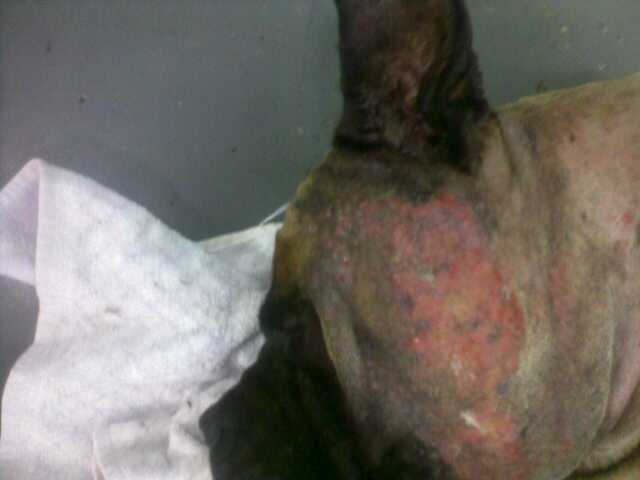
 Aural haematoma – showing swelling of the pinna

Â
Once my dog is treated will I need to bring him/her back to the vet for further treatment?
The sutures will need to be removed 3 weeks after surgery. At this time, a haematoma is usually healed. If an infection is also being treated your veterinarian will also check to make sure that the infection is gone. It is vitally important that the infection is successfully treated to prevent further head shaking which may result in further haematomas.
What happens if your dog does not have surgery?
If a haematoma is left untreated the blood in the ear flap will separate into serum and a clot and will gradually be absorbed over a period of 10 days to 6 weeks. This is an uncomfortable time for your dog and unfortunately some scarring will take place during this process. It also causes a deformity of the ear flap resulting in a “cauliflower ear” which may cause further problems.
Dog ear infections most commonly start as infections of the outer ear, but if neglected they can spread into the middle and inner ear.  this often involves damage to the eardrum.  For this reason, its vital to get your vet to make sure the ear drum is intact before you consider using chemicals in the your dogs ears.  This article “Otitis externa, otitis media, and otitis interna  “  gives a good explanation of how a simple dog ear infection can turn into something much more serious.
Overview
An ear infection, or otitis, is an inflammation of the outer, middle, or inner ear canal. Most frequently, a dog will develop otitis in the outer ear that may worsen and spread into the middle ear. Once in the middle ear canal, the inflammation can move into the inner ear — or, in cases in which the otitis has originated in the middle ear, the infection can instead progress outward to the external ear.
Otitis can be caused by a tremendous array of factors, including fleas, excess liquid in the ear from swimming, autoimmune diseases, skin parasites, and excess wax production. Generally, dogs that develop ear infections have reddened ears that are painful to the touch and ooze a foul-smelling liquid. A punctured eardrum is not an uncommon result of a middle ear infection, and the more severe cases of otitis can lead to partial deafness, lack of balance, nausea and vomiting, and problems with the nerves of the eye.              Clinical Signs
Clinical signs can vary depending on the severity and location of the infection, but typical symptoms of otitis include pain and erythema of the pinna, head shaking and scratching of the ears, and purulent and malodorous exudate coming from the ear canal. The animal may tilt the affected ear downwards and may roll or lean to the affected side. If both sides are affected, the animal may be deaf or ataxic. Some animals may be nauseated, vomit, and have Horner’s syndrome. Any neurological clinical signs indicate significant middle ear or inner ear disease. Symptoms
Ears that are red, painful to the touch, and produce a foul-smelling discharge are symptomatic of otitis. Typically, a dog with an ear infection will scratch and shake the ears or may tilt the affected ear downwards. Animals that are affected more severely may show some neurological signs such as rolling or leaning to one side. If both ears are affected, the animal may be deaf or off balance and uncoordinated. Some animals may be nauseated and vomit. Also, some dogs may get a condition called Horner’s syndrome in which the pupils are sized differently and the nictitating membrane is raised. This condition indicates that a nerve has been affected by the inflammation from the middle ear. Any neurological clinical signs indicate significant middle ear or inner ear disease. Description
Typically, ear infections begin with otitis externa and then progress deeper into the canal to the middle ear. When the inflammation in this region of the ear is chronic, the eardrum may rupture and the infection may spread to the inner ear — or, the infection may begin in the middle ear and progress outward to the external ear. Of the three types of otitis, infections in the inner ear are often the most severe and can lead to partial deafness and neurological problems. In serious cases of otitis, the skin begins to form into folds in which the infection can become trapped, making cleaning and use of topical treatments very difficult. In addition, in cases of otitis interna, the skin will secrete more wax and debris that allows yeast and bacteria to overgrow, causing further disease. Severe inflammation leads to permanent skin thickening, mineralization, and narrowing of the ear canals. Once this occurs, the only viable treatment will be surgical removal of part or all of the ear canal.                Diagnosis
The veterinarian will make a diagnosis based on the clinical signs, physical exam findings, and through the use of several other diagnostic tools. One such tool is cytology, which involves taking a swab of the ears and looking at the material collected under a microscope for the presence of bacteria, yeast, mites, and other substances that might cause an infection. Bacteria and yeast are normally present in low numbers in all dogs’ ears, but a large presence will lead to an ear infection. Skin biopsies may be needed to determine any diseases such as an autoimmune disorder that could cause a skin abnormality affecting the ear. Skin scrapings may be needed to detect mites, tiny parasites that can infect the ears and cause skin diseases. X-rays of the skull can be used to examine the middle and inner ear for signs of disease. In addition, cultures of an infected ear help determine the presence and type of bacteria, as well as antibiotics that are appropriate for treatment. The majority of animals with ear problems have such pain in their ears that they cannot withstand having them examined, cleaned, cultured, or x-rayed without the use of general anesthesia.             Prognosis
With proper treatment, otitis externa usually will resolve within three to four weeks, although it may recur in certain animals. The prognosis is good for otitis media and otitis interna since medical treatment usually is effective. The key for long-term success is correcting or treating the underlying problem that led to the development of the otitis.  Transmission or Cause
Possible causes of otitis externa, which may lead to infections of the middle and inner ear, include excess moisture in the ears due to swimming or bathing; inappropriate treatment or cleaning of the ears such as may be caused by the use of cotton-tipped applicators; excess wax production in the ears; an underlying disease; or a foreign body that is obstructing the ear canal. Additionally, otitis externa can be caused by a host of parasites, including ear mites, mange, fly bites, and fleas. Allergies and illnesses such as autoimmune diseases may also lead to otitis. The most common cause of the development of otitis media and otitis interna is a bacterial infection resulting from otitis externa.  Treatment
After the cause of the otitis has been diagnosed, the veterinarian usually will perform a thorough cleaning of the ear canals while the dog is under sedation or general anesthesia. The use of topical medications, which are placed into the ear canal, is often very beneficial in killing yeast, bacteria, and mites. Oral medications also may be used in conjunction with other treatments to help kill bacteria, yeast, and mites. The veterinarian will teach the dog’s owner how to clean and medicate the ears properly to ensure successful treatment of otitis. The surgical correction of the ear canals may be necessary in cases of severe infection. One type of surgery, called a lateral ear canal resection, allows the ear to drain more easily, decreases the amount of humidity in the ear, and makes topical treatments easier to apply. A total ear canal ablation — a complete removal of the ear canal — is performed on dogs with severe, chronic ear disease that is nonresponsive to medical therapy.           Prevention
Prevention is best accomplished when the veterinarian is able to determine the underlying factors that can lead to development of otitis. Proper cleaning of the ears is also critical. A veterinarian can explain how to clean the ears, as well as how to apply any medications that have been prescribed to treat the otitis.
Dog Ear Infections vary form mild to severe and have many causes. Â To your average owner, these all look the same, but your vet has the skills, experience and equipment to locate the cause and assess the severity of the problem.
Morris Animal Hospital has a great web page showing just what infected, dirty, hairy and blocked ears look like inside.
Dog Vet
Dermatology and Diseases of the EarÂ
Otoscope Case Examples
 |  |  |
| A normal canine eardrum. The eardrum is located 3 inches deep in the canal in large dogs. Note the eardrum (green) and the small amount of wax adhered to the normal tuft of hair that frequently grows next to the eardrum (red).  | Excessive amounts of hair can be normal in breeds such as the Poodle and Bichon Frise. Sometimes the hair causes moisture and wax to be retained in the canal causing an ear infection.  | This large wax plug located deep in the ear canal of a cat caused a good deal of discomfort. It was removed under anesthesia with the video otoscope.  |
 |  |  |
| This Labrador Retriever had severe bilateral otitis and was in excruciating pain. Note the copious amounts of discharge and wax which was subsequently cleansed from the canal with the video otoscope.  | Polyps are sometimes the cause of chronic ear infections. Note how this large polyp is completely obstructing the canal. Polyps cannot be diagnosed and adequately treated until the canal has been thoroughly cleaned.  | This older cocker spaniel has likely had ongoing infections for years. Note the hemorrhage and inflammation. This photo was taken after a tremendous amount of wax and debris were removed. Her ear drum is ruptured but she has taken her first step toward full recovery.  |
 |  |  |
| A young female German Shepard named “Heidi” with a diseased canal lining and ruptured ear drum. She later had surgery to reroute her ear canal which helped prevent future infections & relieved her pain. | A springer spaniel with complete ear drum rupture & otitis media (middle ear infection). Beyond the hair where the ear drum should be, the interior of the middle ear can be seen. This dog needed antibiotics for 6 weeks to resolve her middle ear infection. | After extensive flushing and removal of debris, a normal ear drum is finally visualized in the depths of this ear canal. The catheter being used in this photo is only 1.5 mm in diameter. It is impossible to clean ear canals to this extent without the use of the video ototscope. |
Dog ear infections commonly recur. Â People often blame this on antibiotic resistance or problems with their vet, but the main causes are actually failure to ensure the ear is clean (and stays clean afterward) and failure by owners to put ear drops in the correct location. Â The wikipedia article on dog ear infections has this useful image describing where to put ear drops when medicating your dogs ear.
 Â
 Dog ear infections are common, but there are many possible causes.  I love this article 8 Possible causes of dog ear infection because its simple, accurate, and straight to the point.

Dog Ear Infection
8 Possible Causes Of Dog Ear Infection
Dog ear infection, otitis externa, is a condition of the inflammation of the outer ear canal.
It is a common condition in pets. An estimate of 20% of the dog population is affected by this ear problem.
The structure of a dog’s ear canal differs from human. Human has a horizontal ear canal. A dog has a L-shape ear canal. It is easy for ear wax and moisture to be trap in the vertical section of the dog’s ear canal. Often, the blockage leads to a dog ear infection.
There are many reasons for ear infection in dogs. Knowing the cause of the problem comes in handy when you want to help your dog to get better.
Here are 8 possible causes of dog ear infection:
- Floppy versus Erect Ears
Long floppy ears blowing in the wind makes a dog looks stylish and elegant. But those same floppy ears have a drawback. They block off light and reduce the air circulation within the ears.
Yeast and bacteria multiply quickly in a dark, moist and warm environment. The ear canal of a long, floppy-ear dog is the perfect place. Breeds like Basset Hound, Beagle and Cocker Spaniels often have this ear problem.
- Micro Organisms
Bacteria and yeast are one of the most common factors in dog ear infection.
The most common bacteria associated with ear infections are Staphylococcus Intermedius, Proteus Mirabilis, and Pseudomonas Aeruginosa. Pseudomonas bacteria infections are very resistant and often notoriously difficult to control.
Malassezia Pachydermatis is the yeast found most frequently in association with ear infections. It can cause severe ear and skin infections.
A healthy dog has a balanced flora of yeast and bacteria in the ear canal. But when the ear environment changes due to excess moisture, allergies or hormone disorder, the yeast and bacteria multiply quickly and the ear becomes inflammed.
- Parasites
Ear mites cause intense itching to your dog. A dog’s ear that is infested with ear mites can have thousands of these tiny bugs crawling in the ear canal. As the mites feed and eat into the skin, the ear canal becomes sore and inflamed.
A dog trying to relieve the itch by scratching relentlessly, may cause further damage to the ear flaps.
- Allergies
Underlying allergies sometimes manifest as dog ear infection. A dog can have an allergic reaction to its food, something it inhales, or something it comes in contact with.
It is important to address the allergy in addition to the infection so as to minimize recurring ear inflammation.
- Foreign Bodies
Things that can get into your dog’s ear include insects, twigs, grass seeds, sand, water, pet’s fur … and the list goes on. The foreign object causes irritation. If it travels deeper into the ear, it can cause damage to the ear drum.
- Hematoma
Sometimes a ear infection leads to hematoma in a dog. A dog scratching and shaking its head vigourously to get rid of an itch can break the blood vessels in the ear flap causing bleeding. The ear flap becomes swollen as blood collects within the ear flap. This can be very uncomfortable for the dog.
- Hypothyroidism
Hypothyroidism is the condition where one has inadequate active thyroid hormone. It is the most common hormone imbalance of dogs. Hypothyroidism generally develops in middle aged or elderly dogs.
An imbalance in the thyroid hormone can also lead to dog ear infection.
- Tumors
Ear tumors in dogs can be benign or malignant. They generally affect middle aged or older dogs. Ceruminous gland adenocarcinoma is the most common malignant tumor of the ear canal of dogs, especially cocker spaniels. Other tumors may include papillomas and squamous cell carcinomas.
Some tumors cause pain, ulceration, bleeding and secondary ear infections.
If your pet has dog ear infection, do you know what causes the ear problem?
It may be difficult for a pet guardian to accurately determine the cause of a dog ear infection. So it is advisable to bring your dog to the veterinarian. An otoscopic examination and some lab work would provide a diagnosis of the ear infection.
Once you know the cause of the dog ear infection, you or your vet would be able to apply the appropriate treatment options.
Otitis externa, or inflammation of the external ear canal, is a common condition in dogs and cats. It is characterized by inflammation of the soft tissue components of the external ear canal and is particularly prevalent in dogs with long, floppy ears.Â
 Â
  Â
 Left: as demonstrated by the above ear-model, the ear canal has a vertical and a horizontal component. This predisposes the animal to ear infections as debris must work its way upward rather than straight out.Â
Right: otitis externa with brown exudate Anatomy of the earÂ
The ear of the dog and the cat is composed of three parts: the external ear, the middle ear and the inner ear. Together these components allow the animal to locate a sound and the direction from which it emanates, to orientate the head in relation to gravity and to measure acceleration and rotation of the head.Â
Selective breeding, of dogs in particular, has resulted in a wide variation in relative size and shape of the components of the external ear. Compare, for example, the French Bulldog, the Cocker Spaniel, the German Sheperd Dog, the St. Bernard and the Persian cat. The pinnal shape and carriage, the diameter of the external ear canal, the degree of hair and amount of soft tissue within the external ear canal, and the shape of the skull within which the middle and inner ear lie vary from one breed to another. The external ear canalÂ
An importrant difference between the tissues of healthy ears and infected ears is the appaerance and distribution of the glandular structures. In a healthy ear, sebaceous glands are usually numerous , large and actively secreting. In chronic otitis externa, they are less active and much smaller. The external ear canal may normally contain a small amount of wax that is yellowish-brown in color. Signs of ear disease
Every day we see dogs and cats who have problems with their ears. Signs of these ear problems include:Â
• OdorÂ
• Scratching or rubbing of ears and headÂ
• Discharge in the earsÂ
• Redness or swelling of the ear flap or canalÂ
• Shaking of the head or tilting it to one sideÂ
• Pain around the earsÂ
• Changes in behavior such as depression or irritability Causes of ear diseaseÂ
• Allergies such as atopy or food allergiesÂ
• Parasites - ear mites. Adult mites are large, white and free-moving. the life cycle of the ear mite is three weeks. Mites are characterized by thick, reddish-brown or darkbrown exsudate and crusts in the ear. Ear mites are the single most frequent cause of otitis externa in the cat. Mites are highly contagious.Â
• Bacteria and Yeast infections. Staphylococcus or Streptococcus (Gram-positive bacteria) infections are most frequently associated with acute otitis externa. There is a light-brown, creamy exudate. Gram-negative organisms (Proteus, Pseudomonas, E.Coli) are most frequently observed in chronic otitis externa or recurrent ear disease. There is a yellow exudate. Malassezia canis is a budding yeast: a brown, waxy adherent exudate.Â
• Ticks. The spinous ear tick is found in the external ear canal of dogs and cats. The ear canal become packed with immature ticks. Treatment involves mechanical removal, spraying or dipping the coat with an insecticide.Â
• Foreign bodies, e.g., plant awnsÂ
• TraumaÂ
• Hormonal abnormalities, e.g., hypothyroidismÂ
• The ear environment, e.g., excess moisture and ear anatomyÂ
• Hereditary or immune conditionsÂ
• Tumors TreatmentÂ
The first step in otitis externa is a thorough cleansing of the ear canal followed with irrigation of the canal with a warm antiseptic solution (chlorhexidine or betadine). A warm sterile solution should be used if there is any indication of ruptured membrane.Â
Most topical ear medications are combinations containing one ore more antiparasitics, antibacterial, antimycotic, anti-inflammatory, local anesthetic, ceruminolytic and drying and cleaning agents. Oil or ointment bases help moisturize the skin and are effective in dry, scaly or crusty lesions. Solutions or lotions are most frequently used in exsudative ear lesions.Â
Cotton applicator swabs can be used to clean the inside of the earflap and the part of the ear canal you can see. They should NOT be used farther down in the ear canal since that tends to pack debris in the ear canal, rather than removing it!Â
Chronic otitis externa is usually associated with underlying skin conditions such as seborrhea, hypothyroidism or improperly or neglected ear disease.ar problems caused by a systemic disease such as a hormone abnormality or allergy must include a therapy that treats the whole dog, such as hormonal replacement or allergy testing and hyposensitization (immunotherapy). Surgical treatment
Depending on the severity of the problem, the vertical canal may need to be opened surgically. This enables debris to be removed more effectively. This is done to prevent severe scarring after prolonged specific medical therapy has been ineffective. This procedure is called a lateral ear resection. There are several surgical techniques for this surgery.Â
If the canal becomes so scarred that it is practically closed, ablation may be the final option. In this surgical procedure, the entire ear canal is removed and healthy tissue is allowed to grow in. This procedure is a last resort after severe infection has made effective medical treatment impossible. A specialist is called in for these cases and although surgery is expensive, dogs with chronic severe otitis usually require no further ear treatment for the rest of their lives.Â
 Â
 Aural hematomaÂ
Complication of ear disease:Â aural hematomaÂ
When a dog or cat with uncomfortable ears shakes and scratches vigorously, a blood vessel in the ear flap may rupture. This leads to bleeding into the tissues of the pinna (see above illustration). The usual recommendation is to have the blood clots removed and the ear bandaged and cleaned under anesthesia. If the hematoma is not so big as to occlude the ear canal (thus preventing medication), the option to forgo surgery exists; but without surgery, the ear may scar down into an abnormal appearance. Preventing ear disease
The key to healthy ears is to keep them clean. Check your cat’s and dog’s ears weekly. A slight amount of waxy buildup may be present in normal ears. If your dog swims a lot, has pendulous ears, or a history of ear disease, routine cleaning (often once to three times per week) is recommended. Use the same procedure as described above. Excess hair around the ear can be clipped to allow more air flow. Treat any underlying condition that predisposes your dog to ear problems.Â
Remember, if your dog or cat is showing severe discomfort, the ears have a bad smell, or the ear canals look very abnormal, do not delay in contacting your veterinarian. If your dog has a ruptured or weakened eardrum, some ear cleansers and medications could do more harm than good. f
- Ears aren’t very hairy
- Ears stay clean and smell good without plucking
- The weather is very cold: hair in ears keeps these structures warm
- Ears are sore or infected at the moment: in this case plucking should be done under sedation or anaesthesia, or deferred until the ears have been treated. Â Plucking sore or infected ears can make things worse, and should only be done on the advice of a vet.
Dog Ear Types
There are a variety of different ear shapes and sizes in the canine world. Here are some of the basics with brief descriptions and examples of breeds with each ear shape.

Large in proportion to the head & upright.
Example of breeds with bat ears: Chihuahua & Cardigan Welsh Corgi

Large, upright ears with blunted or rounded tips.
Example of breeds with blunt-tipped ears: Chow Chow & French Bulldog

A small semi-erect ear with a front flap that folds forward nearly to the skull obscuring most of the ear canal.
Example of breeds with button ears: Jack Russell Terrier & Fox Terrier

Specific ear type of the English Toy Terrier breed.

An upright prick ear that folds over slightly at the tip.
Examples of breeds with semiprick ears: Rough Collie & Pitbull

Created by surgically cropping them shortly after birth so they stand up straight. (This is cosmetic surgery)
Example of breeds often found with cropped ears: Doberman & Great Dane

Can be expressed in a variety of shapes & lengths and specifies only that the ear hang down from their junction with the side of the head.
Examples of breeds with drop ears: Basset Hound & Skye Terrier
没有评论:
发表评论